
Featured Collection
Your cart
Featured Collection

PEAK SURGICALS
SKU:PSPS-001
Couldn't load pickup availability
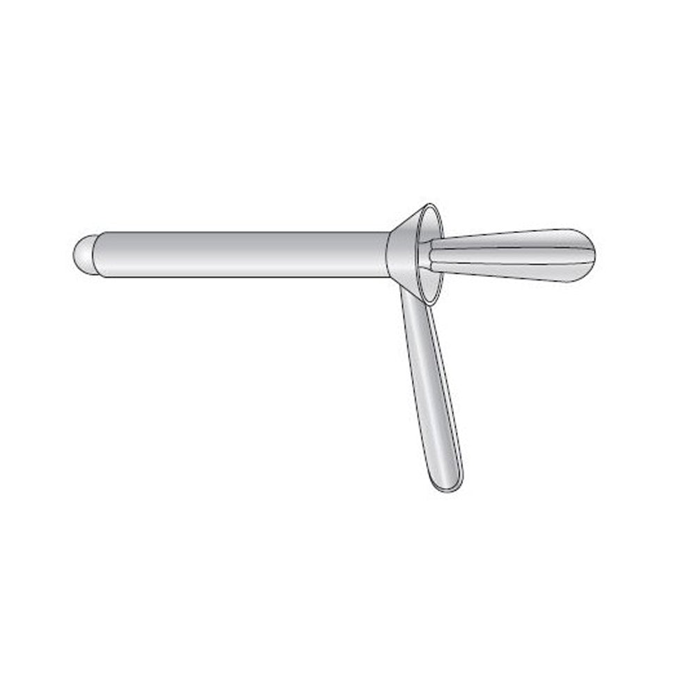
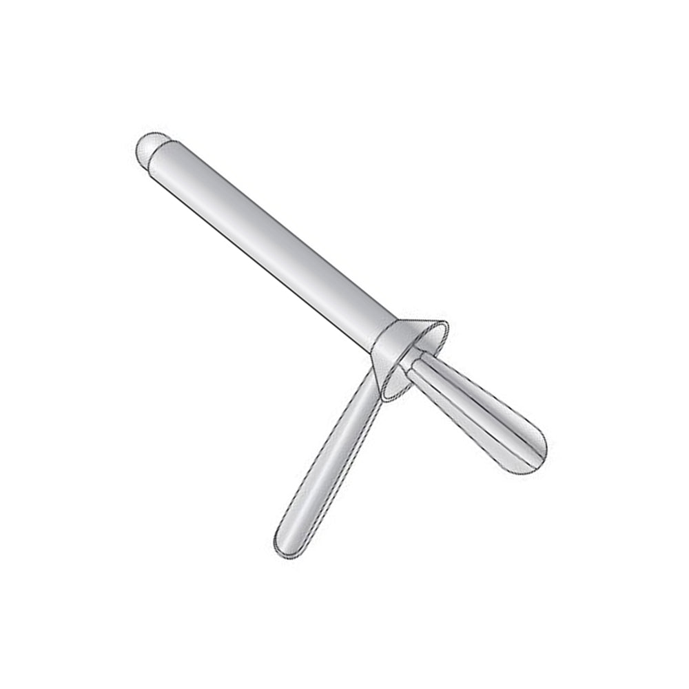
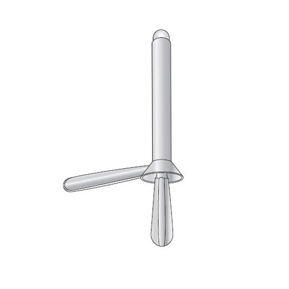
The Kelly Proctoscope from Peak Surgicals (SKU: PSPS-001) is a reusable medical grade stainless steel proctology instrument designed for direct anorectal visualization, rectal canal access, and controlled examination of the distal rectum and anal canal. This product is listed with a 5.5mm size and includes an obturator with a 5.5 long lumen configuration, with each instrument priced at $41.80. The visible design includes a cylindrical proctoscope tube, rounded distal tip profile, removable obturator, fluted obturator handle, lateral handle, proximal viewing flange, smooth internal lumen, and straight tubular body. Colorectal surgeons, general surgeons, proctologists, gastroenterology procedure teams, hospital operating rooms, outpatient surgical clinics, emergency departments, and procurement teams use this instrument during proctoscopy, anorectal examination, hemorrhoid evaluation, rectal bleeding assessment, anal fissure inspection, fistula opening assessment, distal rectal lesion visualization, biopsy access, foreign body assessment, and postoperative anorectal review. The obturator supports smooth insertion, while the lumen provides a direct working channel for illumination, visualization, suction, swab passage, and instrument access.
The Kelly pattern works through a straight cylindrical tube that creates a stable visual and working corridor through the anal canal into the distal rectum. The removable obturator fills the lumen during insertion and presents a rounded leading profile, reducing mucosal catching at the anal verge, dentate line, anal columns, and lower rectal folds. Once the proctoscope is seated, the obturator is withdrawn and the hollow lumen remains available for inspection, illumination, suction, gauze swab passage, biopsy forceps access, or application of topical agents. The proximal flange defines the viewing entrance and gives the clinician a stable edge for manipulation during depth adjustment. The lateral handle provides leverage and orientation control without obstructing the lumen. This is important because proctoscopy requires gradual advancement, rotation, and withdrawal while maintaining the axis of the anal canal. The straight wall preserves the examination channel, while the smooth internal surface supports unobstructed visualization of hemorrhoidal columns, rectal mucosa, bleeding points, fissures, lesions, and postoperative tissue changes.
During proctoscopy, the obturator is inserted with the tube to guide atraumatic entry through the anal canal, then removed to allow direct inspection of the lower rectum. In hemorrhoid evaluation, the lumen allows visualization of internal hemorrhoidal columns, mucosal prolapse, thrombosed changes, bleeding surfaces, and treatment sites after banding or injection therapy. During rectal bleeding assessment, the instrument gives a controlled channel for identifying fissures, inflamed mucosa, ulceration, distal rectal polyps, traumatic injury, or postoperative bleeding. In anal fissure evaluation, the tube supports exposure of the anal canal while the clinician examines the posterior midline, anterior midline, sphincter tone response, and sentinel tag region. During fistula assessment, the proctoscope can help visualize the internal opening, mucosal changes, and drainage site in relation to the dentate line. For biopsy access, the open lumen supports passage of forceps toward visible distal rectal lesions. In postoperative review, it supports inspection after hemorrhoidectomy, fissure surgery, fistula treatment, rectal lesion excision, and anorectal wound care.
The listed 5.5mm size identifies the product variant on the page, while the product description also states a 5.5 long lumen with obturator. In clinical use, the key value of this configuration is controlled access through the anal canal with a lumen that remains stable after obturator removal. The obturator-guided approach is essential because the anal canal has sensitive mucosa, sphincter resistance, vascular hemorrhoidal cushions, and folded rectal lining that can catch against an open tube. The rounded obturator tip allows the tube to advance with a smoother leading surface, then the hollow channel becomes available for examination. The proctoscope tube supports direct evaluation of the distal rectal mucosa, anal columns, hemorrhoid tissue, fissure margins, inflammatory changes, and lesion surfaces. The lateral handle helps the clinician maintain orientation as the instrument is rotated or withdrawn. This controlled entry-and-viewing sequence allows anorectal examination, distal rectal inspection, and minor procedural access through one reusable instrument.
Medical grade stainless steel gives the Kelly Proctoscope the rigidity, corrosion resistance, and surface durability required for repeated proctology use and sterilization workflow. A proctoscope must retain a smooth distal contour, round lumen, stable flange, and secure obturator fit because distortion can interfere with insertion, viewing, or instrument passage. The straight cylindrical tube supports consistent lumen geometry, while the removable obturator must seat cleanly within the working channel before introduction. After use, the instrument is decontaminated, the lumen is brushed or flushed, the obturator is cleaned separately, the handle junction is inspected, and the tube is steam sterilized by autoclave after complete drying. Satin and dull finishes reduce glare from procedure lights, while mirror finish provides a smooth inspection surface after cleaning. The CE marking, ISO-13485 certification, and FDA compliance support hospital purchasing, distributor documentation, and proctology instrument tender files. The listed 1-year warranty, reusable construction, and 30-day return policy support procurement planning for colorectal, general surgery, and outpatient examination sets.
| SKU | PSPS-001 |
|---|---|
| Product Name | Kelly Proctoscope |
| Price | $41.80 per piece |
| Size/Gauge Variants | 5.5mm; with obturator and 5.5 long lumen |
| Instrument Category | Proctology Instruments / Proctoscopes |
| Procedure | Proctoscopy, Anorectal Examination, Hemorrhoid Evaluation, Rectal Bleeding Assessment, Anal Fissure Inspection, Fistula Opening Assessment, Distal Rectal Biopsy Access, Postoperative Anorectal Review |
| Material | Medical Grade Stainless Steel |
| Finish | Satin, Dull, Mirror |
| Sterilization | Steam sterilizable / Autoclave compatible |
| Instrument Classification | Class I |
| Reusable | Yes |
| Certifications | CE, ISO-13485, FDA |
| Warranty | 1 Year |
| MOQ | 1 Piece |
| OEM / Custom Orders | Available |
| After-Sale Service | Return and Replacement |
How does the Kelly Proctoscope compare with a Pratt rectal speculum?
The Kelly Proctoscope provides a cylindrical viewing lumen for direct inspection of the anal canal and distal rectum. A Pratt rectal speculum uses spreading blades to open the anal canal and is more focused on exposure by mechanical dilation. The Kelly pattern is better suited when the clinician wants a stable tubular channel for proctoscopy, rectal bleeding assessment, hemorrhoid visualization, biopsy access, and postoperative inspection. The obturator-guided tube supports smoother insertion before the open lumen is used for examination. A Pratt speculum gives wider separation at the anal verge, but it does not provide the same straight tubular inspection channel into the distal rectum. The Kelly Proctoscope is therefore selected for endoluminal visualization and controlled canal access. In proctology trays, both instruments serve different exposure methods rather than replacing each other.
Why is the obturator important during proctoscope insertion?
The obturator is inserted inside the proctoscope tube before the instrument enters the anal canal. It creates a rounded leading end so the open rim of the tube does not catch on the anal verge, hemorrhoidal cushions, mucosal folds, anal columns, or lower rectal lining. Once the tube is placed, the obturator is withdrawn and the lumen becomes available for direct visualization and instrument passage. This sequence is clinically important because anorectal mucosa is vascular, folded, and sensitive to focal pressure. The Kelly Proctoscope uses the obturator to create a smoother transition from insertion to examination. The fluted obturator handle also allows controlled removal without disturbing the tube position. This makes the instrument practical for hemorrhoid assessment, rectal bleeding evaluation, fissure inspection, and distal rectal lesion review.
What do CE, ISO 13485, and FDA compliance mean for procurement?
CE marking supports conformity documentation for hospitals, clinics, and distributors sourcing reusable surgical instruments. ISO 13485 indicates that manufacturing is managed under a medical device quality management system with controlled production, inspection, and traceability. FDA compliance supports procurement files for buyers sourcing reusable stainless steel instruments for regulated markets. The Kelly Proctoscope is a reusable non-powered proctology instrument, which fits standard purchasing categories for colorectal and general surgery examination tools. These credentials help procurement officers compare the item against tender requirements, internal sourcing policies, and distributor documentation needs. The listed reusable steel construction, 1-year warranty, and 30-day return policy strengthen the ordering record for repeat supply. For colorectal departments and outpatient procedure rooms, the documentation aligns clinical function with regulated purchasing expectations.
How is the lateral handle used during anorectal examination?
The lateral handle is used to guide insertion depth, stabilize the tube, and control rotation during the examination. After obturator-guided placement, the clinician uses the handle to maintain the proctoscope axis while the obturator is removed and the lumen is opened for inspection. Small rotational movements allow inspection of the anterior, posterior, and lateral rectal walls, hemorrhoidal columns, fissure region, and mucosal folds. The handle keeps the operator’s grip outside the viewing channel so illumination, swabs, suction, or biopsy forceps can pass through the lumen. The Kelly Proctoscope benefits from this handle because a tubular instrument must remain steady while the examiner evaluates mucosal detail. During withdrawal, the handle allows controlled stepwise inspection of the distal rectum and anal canal. This control supports diagnostic accuracy and procedural access.
How should the Kelly Proctoscope be cleaned and sterilized after use?
After use, the proctoscope and obturator are decontaminated promptly so blood, mucus, stool residue, lubricant, topical medication, and tissue debris do not dry inside the lumen or around the handle junction. The hollow tube is brushed or flushed through its full length because retained residue can obstruct inspection and compromise reprocessing. The obturator is cleaned separately, including the tip, shaft, and fluted handle. The proximal flange, lateral handle, and distal rim are inspected for smoothness and surface integrity before packaging. Stainless steel construction supports steam sterilization by autoclave after complete cleaning and drying. The Kelly Proctoscope should return to the proctology tray only after the lumen is visually clear, the obturator seats correctly, and all external surfaces are clean. Proper reprocessing preserves insertion control, visualization, and repeated clinical usability.
