제품 정보로 건너뛰기
1 3

PEAK SURGICALS

Salvati Operating Proctoscope – 4 cm x 12/19 cm for Transanal Rectal Surgery

Salvati Operating Proctoscope – 4 cm x 12/19 cm for Transanal Rectal Surgery

SKU(재고 관리 코드):PI-010

정가 $80.08 USD
정가 할인가 $80.08 USD
할인 품절
세금이 포함된 가격입니다. 배송료는 결제 시 계산됩니다.
수량
  • 30 Days Money Back Guarantee.
  • 100% Quality Satisfaction.
  • Medical Grade Steel Reusable.
  • CE-CertificateCE Certified
  • FDA-CertificateFDA Certified
  • iso-certificateISO Certified
전체 세부 정보 보기

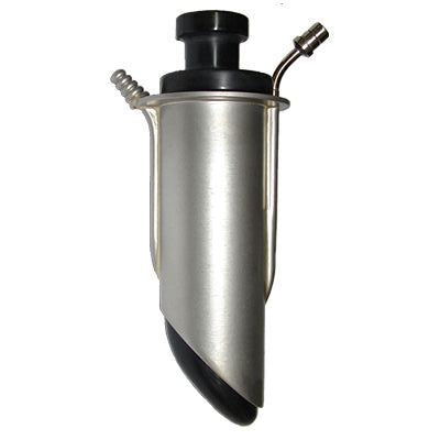
Salvati Operating Proctoscope, SKU PI-008, is a reusable colorectal operating instrument manufactured from German stainless steel for transanal exposure, illuminated rectal visualization, and local excision through a rigid 4 cm working lumen. Two sizes are offered: 4 cm x 12 cm for shorter transanal access and 4 cm x 19 cm for deeper rectal reach. The assembly incorporates a tubular operating barrel, distal beveled opening, proximal flange, fiber-optic light connection, integrated suction pathway, and obturator-style insertion component. The beveled distal profile allows the surgeon to orient the operative window toward a target lesion, while the fiber-optic carrier directs light toward the distal field and the suction port clears blood, fluid, and electrosurgical smoke from the viewing corridor. Colorectal surgeons and general surgeons use this pattern for transanal local excision of rectal adenomas, selected early rectal neoplasms, mucosal or full-thickness lesion resection, biopsy, hemostatic work, and postoperative inspection. The 12 cm version supports conventional long rectal instruments, whereas the 19 cm version provides added reach for more proximal targets and accommodates long laparoscopic-style graspers or electrosurgical instruments. It is suited to colorectal operating rooms, ambulatory surgical units, and hospital procedure suites.

Beveled Operating Lumen, Fiber Optic Light and Suction Mechanism

The Salvati pattern functions as a rigid operating channel rather than a simple diagnostic anoscope. Its 4 cm internal diameter creates enough room for simultaneous visualization and passage of long forceps, electrosurgical pencils, suction, or graspers while maintaining circumferential wall separation around the barrel. The distal end is cut on an oblique bevel so the surgeon can rotate the tube and position the exposed window directly over a rectal lesion. That geometry is clinically important during local excision because the target can be centered within the open face while adjacent mucosa remains supported by the tube wall. A proximal flange provides an external handling reference and limits unintended deep advancement. The fiber-optic light fitting directs illumination along the barrel toward the operative field, reducing dependence on overhead light and improving visualization of mucosal margins, bleeding points, and dissection planes. An integrated suction channel runs alongside the working space and is used to evacuate blood, irrigation fluid, and electrocautery smoke so the line of sight remains clear during resection. The obturator is inserted during introduction to present a smooth leading profile, then removed when the operating lumen is positioned. Depth, rotation, and lesion alignment remain under direct manual control throughout the procedure.

Transanal Local Excision and Rectal Operative Workflow

For transanal local excision, the lubricated proctoscope is introduced with the obturator seated until the planned rectal level is reached, then the obturator is removed and the beveled window is rotated toward the lesion. Fiber-optic illumination is connected before dissection begins, and suction is activated as needed to keep the field clear. The surgeon can mark a circumferential margin around a rectal adenoma or selected early neoplasm, elevate or grasp the lesion, and perform mucosal or full-thickness excision under direct vision. The shorter 12 cm configuration is useful for distal rectal targets that can be reached with long conventional electrosurgical pencils and rectal forceps. The 19 cm configuration is selected when a lesion lies more proximally and additional working depth is required; long laparoscopic-type graspers and electrosurgical tips can be passed through the barrel to reach the operative site. After specimen removal, the lumen permits inspection of the defect, targeted hemostasis, irrigation, suction, and closure with suitable long instruments when indicated. The same rigid access can support biopsy of suspicious lesions and postoperative review of excision sites. Patient selection and oncologic suitability for local excision remain surgical decisions based on lesion stage, location, pathology, mobility, and multidisciplinary treatment planning.

4 cm x 12 cm and 4 cm x 19 cm Size Selection

The two available variants share a 4 cm working diameter but differ in length, which changes reach and instrument handling. The 4 cm x 12 cm version is the compact operating option for lesions in the lower rectum where direct transanal access can be achieved without a long working corridor. Its shorter barrel reduces the distance between the surgeon’s hand and the target, making it practical for conventional long forceps, suction tips, needle holders, and electrosurgical pencils during distal adenoma excision or localized full-thickness resection. The 4 cm x 19 cm version extends the operative channel by 7 cm and is intended for higher rectal targets that cannot be adequately exposed with the shorter device. Greater reach also changes accessory selection, favoring longer laparoscopic-style graspers and cautery tips that maintain control at the distal end of the tube. Both configurations use the same bevel concept to orient the exposed face over the lesion and the same broad lumen to permit simultaneous viewing and instrumentation. The fiber-optic connection serves illumination in either size, while the suction channel removes smoke and fluid during electrosurgery. Hospitals can therefore select the 12 cm model for distal work and the 19 cm model for more proximal transanal local resection while retaining a common operating technique.

German Stainless Steel Reprocessing and Procurement Standards

German stainless steel provides the rigidity needed to preserve the 4 cm operating lumen during insertion, rotation, and sustained rectal wall retraction, while supporting corrosion resistance through repeated decontamination and steam sterilization. A smooth polished finish reduces drag at the patient-contact surfaces and helps sterile-processing staff remove blood, mucus, lubricant, char, and tissue debris from the barrel and bevel. Reprocessing begins promptly after use with separation of removable components, flushing of the suction pathway, brushing of the working lumen, cleaning around the fiber-optic connection, thorough rinsing, complete drying, and inspection for retained soil or mechanical damage. The suction channel requires particular attention because coagulum and electrosurgical residue can obstruct flow if not cleared before sterilization. The optical interface should remain clean and undamaged so light transmission is not reduced during the next case. Reusable stainless components can be processed through a validated hospital steam-autoclave cycle appropriate to the device. CE marking supports applicable conformity documentation, while ISO 13485 identifies the medical-device quality-management framework used for controlled manufacturing, traceability, and documentation. FDA-compliant procurement in the United States requires the appropriate device, establishment, labeling, and import records for the transaction. Peak Surgicals supports institutional ordering with a one-year warranty, single-piece MOQ, OEM or private-label supply, and return or replacement service.

SKU PI-010
Product Name Salvati Operating Proctoscope
Price $72.80 USD
Size/Gauge Variants 4 cm x 12 cm; 4 cm x 19 cm
Instrument Category Proctology Instruments / Operating Proctoscopes
Procedure Transanal local excision, rectal adenoma resection, selected early rectal lesion excision, biopsy, hemostasis and postoperative rectal inspection
Material German stainless steel
Finish Mirror-polished stainless-steel finish
Sterilization Reusable and compatible with validated steam-autoclave processing
Instrument Classification Reusable rigid colorectal operating proctoscope
Reusable Yes
Certifications CE marked, ISO 13485 quality system, FDA-compliant procurement documentation
Warranty 1 year
MOQ 1 piece
OEM / Custom Orders Available for hospitals, distributors and private-label procurement
After-Sale Service Return and replacement support

How does the Salvati Operating Proctoscope differ from a Kelly Proctoscope?
The Salvati Operating Proctoscope differs from a Kelly Proctoscope in both working diameter and operative intent. The Salvati pattern uses a 4 cm barrel that creates substantially more room for long forceps, electrosurgical instruments, suction, and local excision maneuvers. Its distal bevel can be rotated so the open face is positioned directly over a rectal lesion. It also incorporates fiber-optic illumination and a suction pathway to maintain visibility during operative work. A Kelly Proctoscope is generally narrower and is used primarily for diagnostic examination of the anal canal and distal rectum. The Salvati design is therefore selected when direct transanal resection or broader instrument access is required rather than inspection alone.

When should the 4 cm x 12 cm or 4 cm x 19 cm version be selected?
The Salvati Operating Proctoscope is available in 4 cm x 12 cm and 4 cm x 19 cm configurations. The 12 cm model is selected for lower rectal lesions that can be exposed through a shorter transanal working corridor. Its reduced length permits direct handling of long conventional rectal forceps, suction tips, and electrosurgical pencils close to the operative field. The 19 cm version is intended for more proximal rectal targets where an additional 7 cm of reach is needed. That longer barrel is better matched to laparoscopic-style graspers and extended electrosurgical tips used during local excision. Both sizes retain the same 4 cm diameter, so the key selection factor is lesion height and required instrument reach rather than lumen width.

What do CE, ISO 13485 and FDA requirements mean for institutional procurement?
The Salvati Operating Proctoscope is supplied within Peak Surgicals' CE and ISO 13485 procurement framework. CE marking supports applicable medical-device conformity documentation for markets where that marking is required. ISO 13485 identifies the quality-management system used for controlled manufacturing, traceability, documentation, and supplier processes. FDA procurement context for United States buyers concerns the applicable device, establishment, labeling, import, and distribution records associated with the transaction. Hospitals and distributors should retain these records within vendor qualification and purchasing files. These frameworks serve different regulatory functions and should be reviewed independently during institutional sourcing.

How are the bevel, fiber-optic fitting and suction channel used intraoperatively?
The principal intraoperative control features are the beveled distal opening, fiber-optic fitting, suction pathway, and external barrel orientation. After insertion, the Salvati Operating Proctoscope is rotated until the bevel frames the target lesion within the working window. The light connection is coupled to a compatible source so illumination reaches the distal rectal field without occupying the central operating space. Suction is used during electrosurgical dissection to remove smoke, blood, and irrigation fluid that would otherwise obscure the margin or resection plane. The surgeon then controls depth and rotational alignment manually while long instruments pass through the 4 cm lumen. There is no ratchet or self-retaining expansion mechanism because exposure is maintained by the rigid barrel itself.

How should hospitals sterilize and manage the Salvati Operating Proctoscope?
Hospitals should reprocess the Salvati Operating Proctoscope as a reusable colorectal surgical instrument after every case. The working lumen, beveled edge, fiber-optic interface, and suction pathway should be cleaned promptly so blood, mucus, char, and coagulum do not dry inside the device. The suction channel requires flushing and inspection before sterilization because retained debris can obstruct flow during the next procedure. After rinsing and complete drying, staff should inspect the barrel for deformation and confirm that the optical and suction connections remain patent and undamaged. Reusable stainless components can then enter a validated steam-autoclave cycle appropriate to the instrument and facility protocol. Procurement teams should retain SKU PI-008, size selection, warranty, regulatory records, and service history so the 12 cm and 19 cm variants remain traceable in inventory.