Το καλάθι σας είναι κενό
Συνέχιση αγορώνΈχετε λογαριασμό;
Συνδεθείτε για ταχύτερη ολοκλήρωση των αγορών σας.

Επιλεγμένη Συλλογή
Το καλάθι σας
Το καλάθι σας είναι κενό
Συνέχιση αγορώνΈχετε λογαριασμό;
Συνδεθείτε για ταχύτερη ολοκλήρωση των αγορών σας.
Επιλεγμένη Συλλογή

PEAK SURGICALS
SKU:PI-003
Δεν ήταν δυνατή η φόρτωση της διαθεσιμότητας παραλαβής


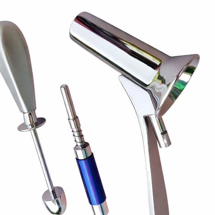
Το πρωκτοσκόπιο Kelly με πηγή φωτός, SKU PSPS-002, είναι ένα επαναχρησιμοποιούμενο άκαμπτο πρωκτολογικό όργανο εξέτασης κατασκευασμένο από γερμανικό ανοξείδωτο χάλυβα για άμεση οπτικοποίηση και ελεγχόμενη πρόσβαση στον πρωκτικό σωλήνα και το άπω ορθό. Η διαμόρφωση εργασίας 22 mm διαμέτρου x 90 mm συνδυάζει σώμα σωληνοειδούς σκοπίου, αφαιρούμενο φραγή, πλευρική λαβή λειτουργίας, ανοιγόμενο εγγύς άνοιγμα προβολής και αποσπώμενο συγκρότημα φωτισμού. Κατά την εισαγωγή, ο φραγής καταλαμβάνει τον αυλό και παρουσιάζει ένα λείο στρογγυλεμένο μπροστινό προφίλ· μετά την τοποθέτηση, αποσύρεται ώστε ο εσωτερικός αγωγός να μπορεί να χρησιμοποιηθεί για φωτισμένη επιθεώρηση και διέλευση κατάλληλων μεγέθους στυλεών, αναρροφητικών άκρων, λαβίδων βιοψίας ή εξαρτημάτων θεραπείας. Η πλευρική λαβή επιτρέπει ελεγχόμενη περιστροφή και απόσυρση κατά τη διάρκεια περιφερικής αξιολόγησης του βλεννογόνου, ενώ το εξάρτημα φωτός κατευθύνει τον φωτισμό κατά μήκος του διαδρόμου εξέτασης. Χειρουργοί παχέος εντέρου, γενικοί χειρουργοί, πρωκτολόγοι και ομάδες γαστρεντερολογικών επεμβάσεων χρησιμοποιούν αυτό το σχέδιο για πρωκτοσκόπηση, αλγόνοψη, αξιολόγηση εσωτερικών αιμορροΐδων, αξιολόγηση ορθικής αιμορραγίας, επιθεώρηση πρωκτικών ρωγμών, εντοπισμό συριγγίου, εξέταση αλγίων του άπω ορθού, στοχευμένη πρόσβαση σε βιοψία και μετεγχειρητική επανεξέταση μετά από αιμορροϊδεκτομή, συριγγιοτομή, χειρουργική επέμβαση ρωγμής ή τοπικές πρωκτοορθικές επεμβάσεις. Προορίζεται για νοσοκομεία, κλινικές παχέος εντέρου, κέντρα χειρουργικής επέμβασης ημερήσιας νοσηλείας και χειρουργεία ή αίθουσες επεμβάσεων.
Το σχέδιο Kelly χρησιμοποιεί έναν ευθύ άκαμπτο σωλήνα για να διατηρήσει έναν καθορισμένο διάδρομο εξέτασης 22 mm μέσω του πρωκτικού σωλήνα. Η κυλινδρική του γεωμετρία εμποδίζει τους γύρω τοίχους να συμπτυχθούν στην οπτική οδό, ενώ ο κλινικός γιατρός επιθεωρεί τον αυλό υπό άμεσο φωτισμό. Ο αφαιρούμενος φραγής περνάει μέσα από το σώμα του σκοπίου πριν από την εισαγωγή και κλείνει το άπω άνοιγμα με ένα στρογγυλεμένο περίγραμμα, μειώνοντας την εμπλοκή του βλεννογόνου σε εκτεθειμένο άκρο σωλήνα κατά την προώθηση. Μόλις το όργανο φτάσει στο επιθυμητό βάθος, ο φραγής αφαιρείται για να εκτεθεί πλήρως ο αυλός για επιθεώρηση και διέλευση εξαρτημάτων. Το διασταλμένο εγγύς άνοιγμα σχηματίζει μια ευρεία είσοδο στον αγωγό εργασίας και βοηθά τον χειριστή να διατηρεί τον προσανατολισμό του κατά την προβολή του φωτισμένου πεδίου. Μια πλευρική λαβή προβάλλει μακριά από τον οπτικό άξονα, επιτρέποντας στον χειρουργό να περιστρέφει, να σταθεροποιεί, να προωθεί ή να αποσύρει το όργανο χωρίς να εμποδίζει το εγγύς άνοιγμα. Το αποσπώμενο συγκρότημα πηγής φωτός συνδυάζει τον φωτισμό με την οδό εξέτασης, ώστε το χρώμα του βλεννογόνου, η αγγειακή αλλαγή, τα σημεία αιμορραγίας, η εξέλκωση και οι ορατές αλλοιώσεις να μπορούν να αξιολογηθούν με βελτιωμένη οπτική ευκρίνεια. Επειδή η συσκευή βασίζεται σε έναν σταθερό σωληνοειδή αυλό αντί για επεκτεινόμενες λεπίδες, η έκθεση παραμένει σταθερή κατά την περιστροφή και τη σταδιακή απόσυρση.
Για διαγνωστική πρωκτοσκόπηση, το συναρμολογημένο όργανο λιπαίνεται με τον οβουράτορα τοποθετημένο μέσα στον σωλήνα και εισάγεται μέσω του πρωκτικού σωλήνα υπό ελεγχόμενη πίεση. Αφού τοποθετηθεί το τμήμα εργασίας των 90 mm, ο οβουράτορας αφαιρείται και προσαρμόζεται το εξάρτημα φωτισμού ώστε ο χειριστής να μπορεί να επιθεωρήσει τον σωλήνα, την περιοχή της οδοντωτής γραμμής, τα αιμορροϊδικά μαξιλάρια και τον προσβάσιμο βλεννογόνο του άπω ορθού. Κατά την αξιολόγηση των εσωτερικών αιμορροΐδων, η σταδιακή περιστροφή και απόσυρση επιτρέπουν την περιφερική επανεξέταση της πρόπτωσης, της συμφόρησης, της ευθρυψίας, της ενεργού αιμορραγίας, της εξέλκωσης ή των αλλαγών των ιστών μετά από θεραπεία στο ιατρείο. Σε ασθενείς που παρουσιάζουν αιμορραγία στο κατώτερο ορθό, ο σταθερός αυλός βοηθά στον εντοπισμό των βλαβών του βλεννογόνου του άπω ορθού, τραυματικών βλαβών, μετεγχειρητικών θέσεων ή αιμορροϊδικού ιστού που αιμορραγεί. Για ύποπτο συρίγγιο, η άμεση οπτικοποίηση μπορεί να υποστηρίξει την αναγνώριση ενός εσωτερικού ανοίγματος πριν από την ανίχνευση ή τον οριστικό χειρουργικό σχεδιασμό. Ο αγωγός επιτρέπει επίσης στοχευμένη στυλεοληψία, αναρρόφηση ή βιοψία μιας ορατής ανωμαλίας με λεπτά εξαρτήματα που ταιριάζουν στον αυλό. Μετά από αιμορροϊδεκτομή, συριγγιοτομή, χειρουργική επέμβαση ρωγμής ή τοπική εκτομή ορθού, οι ομάδες χειρουργών παχέος εντέρου και γενικών χειρουργών μπορούν να χρησιμοποιήσουν το πρωκτοσκόπιο για να αξιολογήσουν την επούλωση, την υπολειπόμενη αιμορραγία, τον σχηματισμό ουλών, τη στενωτική αλλαγή ή την ανωμαλία του βλεννογόνου σε εξωτερικές ή ενδονοσοκομειακές επισκέψεις.
Η διαμόρφωση 22 mm x 90 mm επιλέγεται όταν ο κλινικός στόχος απαιτεί ένα άκαμπτο ανορθικό κανάλι προβολής με μεγαλύτερη εμβέλεια από ένα κοντό πρωκτοσκόπιο, ενώ παραμένει συμπαγές για τις καθημερινές πρωκτολογικές εργασίες. Το διαμέτρημα 22 mm παρέχει χώρο για άμεση επιθεώρηση και περιορισμένη διέλευση εξαρτημάτων χωρίς τη χρήση της ευρύτερης έκθεσης που δημιουργείται από ένα δικατομικό ορθικό κολποδιαστολέα. Το μήκος εργασίας των 90 mm επιτρέπει την εξέταση πέρα από τον άμεσο πρωκτικό σωλήνα στο κατώτερο ορθό, υποστηρίζοντας την αξιολόγηση της αιμορροϊδικής νόσου, των πηγών αιμορραγίας, των άπω βλαβών και του μετεγχειρητικού ιστού. Το σώμα του σκοπίου είναι το κύριο εξάρτημα πρόσβασης και διατηρεί τον κυκλικό διάδρομο εξέτασης καθ' όλη τη διάρκεια της περιστροφής. Ο οβουράτορας είναι το εξάρτημα εισαγωγής· το λείο στρογγυλεμένο άκρο του προστατεύει το ανοιχτό άπω σωλήνα κατά την είσοδο και αφαιρείται όταν αρχίζει η οπτικοποίηση. Η πλευρική λαβή παρέχει έλεγχο κατεύθυνσης κατά την προώθηση, σταθεροποίηση, περιφερική περιστροφή και σταδιακή απόσυρση. Το διασταλμένο εγγύς άνοιγμα διατηρεί την είσοδο εργασίας προσβάσιμη για προβολή και μικρά βοηθητικά όργανα. Το αποσπώμενο συγκρότημα φωτισμού παρέχει εστιασμένο φως κατά μήκος του αυλού, κάτι που είναι ιδιαίτερα σημαντικό κατά τη διαφοροποίηση αγγειακών, φλεγμονωδών, τραυματικών ή μετεγχειρητικών αλλαγών. Μαζί, αυτά τα εξαρτήματα σχηματίζουν μια επαναχρησιμοποιήσιμη διαγνωστική διαμόρφωση κατάλληλη για τυπικές ροές εργασίας εξέτασης του πρωκτικού σωλήνα σε ενήλικες.
Ο γερμανικός ανοξείδωτος χάλυβας παρέχει την ακαμψία που απαιτείται για τη διατήρηση της σωληνοειδούς γεωμετρίας κατά την επανειλημμένη κλινική χρήση, προσφέροντας παράλληλα αντοχή στη διάβρωση υπό επικυρωμένους κύκλους καθαρισμού και αποστείρωσης με ατμό. Η γυαλισμένη σαν καθρέφτης επιφάνεια δημιουργεί μια λεία εξωτερική και εσωτερική επιφάνεια που μειώνει την τριβή κατά την εισαγωγή και υποστηρίζει την απομάκρυνση αίματος, βλέννας, λιπαντικού και άλλων βιολογικών ρύπων κατά την απολύμανση. Η επανεπεξεργασία ξεκινά αμέσως μετά τη χρήση με τον διαχωρισμό του οβουράτορα και του αποσπώμενου εξαρτήματος φωτισμού από το κύριο σώμα σύμφωνα με το πρωτόκολλο της εγκατάστασης, ακολουθούμενη από ενδελεχή καθαρισμό του σωλήνα, των αρμών, των επιφανειών επαφής και των προσβάσιμων αγωγών. Το όργανο στη συνέχεια ξεπλένεται, στεγνώνεται, επιθεωρείται οπτικά, συσκευάζεται και αποστειρώνεται σε επικυρωμένο κύκλο αυτόκαυστου κατάλληλο για επαναχρησιμοποιήσιμα χειρουργικά εργαλεία από ανοξείδωτο χάλυβα. Η επιθεώρηση πρέπει να περιλαμβάνει το άπω χείλος, τον αυλό, τη διασταύρωση της λαβής, την εφαρμογή του οβουράτορα και την περιοχή σύζευξης φωτός, διότι η παραμόρφωση ή τα υπολείμματα μπορούν να παρεμποδίσουν την εξέταση ή την επανεπεξεργασία. Η σήμανση CE υποστηρίζει την ισχύουσα τεκμηρίωση συμμόρφωσης, ενώ το ISO 13485 παρέχει ένα πλαίσιο διαχείρισης ποιότητας ιατροτεχνολογικών προϊόντων για ελεγχόμενη κατασκευή και ιχνηλασιμότητα. Για αγορές στις Ηνωμένες Πολιτείες, η τεκμηρίωση προμηθειών συμβατή με τον FDA πρέπει να διατηρείται εντός της διαδικασίας πιστοποίησης του νοσοκομείου, του διανομέα ή του εισαγωγέα. Η επαναχρησιμοποιήσιμη κατασκευή υποστηρίζει την κεντρική αποστείρωση, τον έλεγχο αποθεμάτων και την επανειλημμένη κλινική χρήση.
| SKU | PI-002 |
|---|---|
| Όνομα Προϊόντος | Πρωκτοσκόπιο Kelly με Πηγή Φωτός |
| Τιμή | 64.00 USD |
| Παραλλαγές Μεγέθους/Διαμετρήματος | Διάμετρος 22 mm x μήκος εργασίας 90 mm, PSPS-002 |
| Κατηγορία Οργάνου | Πρωκτολογικά Όργανα / Πρωκτοσκόπια |
| Διαδικασία | Πρωκτοσκόπηση, αλγόνοψη, αξιολόγηση αιμορροΐδων, αξιολόγηση αιμορραγίας ορθού, επιθεώρηση πρωκτικής ρωγμής, αξιολόγηση συριγγίου, εξέταση βλαβών του άπω ορθού και μετεγχειρητική επανεξέταση πρωκτοορθού |
| Υλικό | Γερμανικός ανοξείδωτος χάλυβας |
| Φινίρισμα | Φινίρισμα γυαλιστερό σαν καθρέφτης |
| Αποστείρωση | Επαναχρησιμοποιήσιμο, αποστειρώσιμο σε αυτόκαυστο και συμβατό με επικυρωμένα πρωτόκολλα αποστείρωσης με ατμό |
| Κατηγοριοποίηση Οργάνου | Επαναχρησιμοποιήσιμο άκαμπτο πρωκτολογικό όργανο εξέτασης |
| Επαναχρησιμοποιήσιμο | Ναι |
| Πιστοποιήσεις | Σήμανση CE, σύστημα ποιότητας ISO 13485, τεκμηρίωση προμηθειών συμβατή με τον FDA |
| Εγγύηση | 1 έτος |
| Ελάχιστη ποσότητα παραγγελίας (MOQ) | 1 τεμάχιο |
| Παραγγελίες OEM / Προσαρμοσμένες | Διαθέσιμες για νοσοκομεία, διανομείς και προμήθειες ιδιωτικής ετικέτας |
| Εξυπηρέτηση Μετά την Πώληση | Υποστήριξη επιστροφής και αντικατάστασης |
Πώς διαφέρει το πρωκτοσκόπιο Kelly με πηγή φωτός από ένα ορθικό κολποδιαστολέα Pratt;
Το πρωκτοσκόπιο Kelly με πηγή φωτός διαφέρει από έναν ορθικό κολποδιαστολέα Pratt στον τρόπο με τον οποίο δημιουργεί έκθεση. Ο σχεδιασμός Kelly χρησιμοποιεί έναν σταθερό κυλινδρικό αυλό που διατηρεί έναν σταθερό διάδρομο προβολής 22 mm, ενώ το σχέδιο Pratt χρησιμοποιεί δύο λεπίδες που ανοίγουν πλευρικά μετά την εισαγωγή. Ο φραγής Kelly κλείνει τον σωλήνα κατά την εισαγωγή και αφαιρείται πριν από την επιθεώρηση, ενώ το όργανο Pratt εισάγεται με τις λεπίδες του κλειστές και στη συνέχεια επεκτείνεται. Ένας άκαμπτος σωληνοειδής αγωγός είναι καταλληλότερος για περιφερική πρωκτοσκοπική προβολή και διέλευση λεπτών εξαρτημάτων μέσω ενός καθορισμένου καναλιού. Ο σχεδιασμός Pratt επιλέγεται όταν απαιτείται ρυθμιζόμενη πλευρική διεύρυνση του πρωκτικού σωλήνα για εξέταση ή χειρουργική πρόσβαση. Για φωτισμένη επιθεώρηση του άπω ορθού, η διαμόρφωση Kelly παρέχει έναν σταθερό οπτικό διάδρομο χωρίς μηχανισμό ανοίγματος λεπίδας.
Γιατί επιλέγεται η διαμόρφωση 22 mm x 90 mm για την πρωκτοορθική εξέταση;
Το πρωκτοσκόπιο Kelly με πηγή φωτός έχει διαμόρφωση 22 mm διαμέτρου x 90 mm μήκους εργασίας για την τακτική πρωκτοορθική εξέταση ενηλίκων. Το διαμέτρημα των 22 mm παρέχει επαρκή χώρο για άμεση προβολή, πρόσβαση αναρρόφησης, στυλεοληψία και λεπτή βιοψία, διατηρώντας ένα σταθερό κυκλικό πεδίο. Η εμβέλεια των 90 mm εκτείνεται πέρα από μια σύντομη πρωκτοσκοπική εξέταση και επιτρέπει την επιθεώρηση του προσβάσιμου βλεννογόνου του κατώτερου ορθού. Κατά την αξιολόγηση αιμορροΐδων, αυτή η διαμόρφωση υποστηρίζει την περιφερική αξιολόγηση του πρωκτικού σωλήνα, της περιοχής της οδοντωτής γραμμής και των εσωτερικών αιμορροϊδικών μαξιλαριών. Κατά την αξιολόγηση αιμορραγίας ορθού ή βλαβών του άπω ορθού, το μήκος εργασίας επιτρέπει στον χειρουργό να προχωρήσει στο κατώτερο ορθό πριν προβεί σε ελεγχόμενη απόσυρση. Αυτό το μέγεθος επιλέγεται επομένως όταν η προγραμματισμένη ροή εργασίας απαιτεί τόσο την οπτικοποίηση του πρωκτικού σωλήνα όσο και περιορισμένη εμβέλεια του άπω ορθού μέσω ενός άκαμπτου πρωκτοσκοπίου.
Τι σημαίνουν οι απαιτήσεις προμηθειών CE, ISO 13485 και FDA για αυτό το όργανο;
Το πρωκτοσκόπιο Kelly με πηγή φωτός παρέχεται στο πλαίσιο προμηθειών CE και ISO 13485 της Peak Surgicals. Η σήμανση CE αφορά την ισχύουσα τεκμηρίωση συμμόρφωσης για τη διανομή ιατροτεχνολογικών προϊόντων σε σχετικές αγορές. Το ISO 13485 προσδιορίζει το σύστημα διαχείρισης ποιότητας που χρησιμοποιείται για την ελεγχόμενη κατασκευή, τεκμηρίωση και ιχνηλασιμότητα ιατροτεχνολογικών προϊόντων. Το πλαίσιο προμηθειών του FDA για τους αγοραστές των Ηνωμένων Πολιτειών περιλαμβάνει τη ρύθμιση, την εγκατάσταση, την επισήμανση, την εισαγωγή και την τεκμηρίωση της συσκευής που ισχύει για τον προμηθευτή και την αγορά προορισμού. Τα νοσοκομεία και οι διανομείς θα πρέπει να διατηρούν αυτά τα αρχεία ως μέρος της διαδικασίας πιστοποίησης προμηθευτών και της τεκμηρίωσης αγορών. Αυτά τα πλαίσια υποστηρίζουν την επισκόπηση των προμηθειών, αλλά εξυπηρετούν διαφορετικές ρυθμιστικές λειτουργίες και δεν πρέπει να αντιμετωπίζονται ως ανταλλάξιμες πιστοποιήσεις.
Πώς χρησιμοποιούνται ο οβουράτορας, η λαβή και το συγκρότημα φωτισμού κατά τη διάρκεια της διαδικασίας;
Το κύριο στοιχείο ελέγχου εισαγωγής είναι ο αφαιρούμενος οβουράτορας, ο οποίος τοποθετείται μέσα στο πρωκτοσκόπιο πριν από την εισαγωγή. Το στρογγυλεμένο άπω προφίλ του καλύπτει την ανοιχτή άκρη του σωλήνα και παρέχει μια λεία επιφάνεια καθώς το συγκρότημα περνάει μέσα από τον πρωκτικό σωλήνα. Αφού φτάσει στο επιθυμητό βάθος, ο οβουράτορας αποσύρεται για να εκτεθεί ο αυλός για εξέταση. Η πλευρική λαβή δίνει τότε στον χειριστή τη δυνατότητα σταθεροποίησης, περιστροφής, διόρθωσης κατεύθυνσης και σταδιακής απόσυρσης χωρίς να μπλοκάρει το εγγύς άνοιγμα προβολής. Το αποσπώμενο συγκρότημα φωτισμού κατευθύνει το φως μέσω του άκαμπτου διαδρόμου, έτσι ώστε οι επιφάνειες του βλεννογόνου και τα σημεία αιμορραγίας να παραμένουν ορατά κατά την κίνηση. Το πρωκτοσκόπιο Kelly με πηγή φωτός βασίζεται επομένως στην τοποθέτηση του οβουράτορα, στον έλεγχο της λαβής και στην φωτιζόμενη οπτικοποίηση και όχι σε μηχανισμό καστάνιας, άξονα ή επέκτασης λεπίδας.


